

Understanding the differences in normal vs abnormal shoulder MRI can significantly impact diagnosis and treatment strategies. A normal MRI shows clear and well-defined structures, while an abnormal MRI often reveals tears, inflammation, or other issues that require attention.
Recognizing these distinctions helps both patients and healthcare providers navigate the complexities of shoulder health. Let’s delve into the key indicators that differentiate a normal shoulder MRI from one that suggests a problem, enhancing your knowledge and awareness in this critical area of medical imaging.

Normal vs Abnormal Shoulder MRI: Understanding the Differences
Shoulder MRI scans are vital tools used by doctors to view the internal structures of the shoulder joint. They help in diagnosing various conditions that can affect the shoulder, such as tears, inflammation, or other abnormalities. Understanding what a normal versus an abnormal MRI looks like can empower patients to engage in informed discussions with their healthcare providers. In this article, we’ll delve deep into the characteristics of normal and abnormal shoulder MRI results, the common conditions depicted, and how these findings influence treatment options.
What Is a Shoulder MRI?
A shoulder MRI (Magnetic Resonance Imaging) uses powerful magnets and radio waves to create detailed images of the shoulder’s structures. It provides a non-invasive way to visualize soft tissues, such as muscles, tendons, and ligaments, as well as cartilage and bones.
How Does an MRI Work?
– **Magnetic Field**: The MRI machine generates a strong magnetic field that aligns the water molecules in the body.
– **Radio Waves**: Once aligned, radio waves are sent through the body, causing the molecules to produce signals.
– **Image Creation**: Those signals are collected and converted into images by a computer.
Understanding this process helps demystify how doctors can see what’s happening inside the shoulder without needing surgery.
Normal Shoulder MRI Characteristics
When looking at a normal shoulder MRI, physicians expect to identify several key features:
- Intact Rotator Cuff: The rotator cuff is a group of muscles and tendons that stabilize the shoulder. In a healthy MRI, these structures appear continuous and free from tears.
- Normal Joint Space: The shoulder joint space should be well-defined, indicating proper cartilage health.
- No Fluid Accumulation: There should be no excess fluid around the joint or within the soft tissues.
- Intact Labrum: The labrum, a fibrocartilaginous structure, should be normal in shape and without fraying.
- Well-Defined Bones: The humeral head, glenoid, and other bones should have a smooth surface and no signs of erosion.
These characteristics are essential for a doctor’s assessment of shoulder health. A normal MRI indicates that everything looks healthy and functioning as it should.
Common Abnormal Findings in Shoulder MRI
When a shoulder MRI shows abnormalities, it can indicate various conditions. Here are some common findings:
Torn Rotator Cuff
– **Description**: A tear in one or more of the rotator cuff tendons.
– **MRI Appearance**: MRI will typically show fluid in the tear, leading to a discontinuity in the tendon.
– **Symptoms**: Pain, weakness, and limited range of motion in the arm.
Shoulder Impingement
– **Description**: This condition occurs when the rotator cuff tendons get pinched during shoulder movements.
– **MRI Appearance**: You may observe swelling of the tendons and abnormal positioning of the acromion (the bony part on the top of the shoulder).
– **Symptoms**: Pain during overhead activities and a feeling of weakness.
Labral Tears
– **Description**: A tear in the ring of cartilage (labrum) surrounding the shoulder socket.
– **MRI Appearance**: A labral tear may appear as a frayed edge or a detachment from the bone.
– **Symptoms**: Clicking or popping sensation, along with shoulder pain.
Arthritis
– **Description**: Inflammation of the shoulder joint can be caused by wear and tear, known as osteoarthritis, or autoimmune disorders like rheumatoid arthritis.
– **MRI Appearance**: MRI may show joint space narrowing, bone spurs, and cyst formation.
– **Symptoms**: Stiffness, swelling, and pain that gets worse with activity.
Bone Fractures
– **Description**: Breaks in the bones of the shoulder due to trauma or injury.
– **MRI Appearance**: Fractures may be visible as a break in the bone continuity.
– **Symptoms**: Severe pain, inability to move the arm, and visible deformity.
Interpreting MRI Results: What to Expect
Understanding how to interpret MRI results can be challenging. Here’s what you should know:
- Consult with a Specialist: Always consult with your physician or a radiologist who can explain the findings in detail.
- Understand Terminology: Familiarize yourself with common MRI terms your doctor may use, such as “tendonopathy,” “edema,” or “lesion.”
- Consider Follow-Up Tests: Sometimes, an MRI isn’t enough to provide a complete picture, and your doctor may recommend additional imaging or tests.
Reading MRI results involves both the images and your medical history. Any findings need to be interpreted in the context of your symptoms and physical examination results.
Importance of MRI in Diagnosis and Treatment
MRI plays a crucial role in diagnosing shoulder issues and planning treatment. Here’s how:
- Accurate Diagnosis: An MRI provides detailed images that help identify the exact cause of shoulder pain, leading to a more accurate diagnosis.
- Guiding Treatment Plans: Based on the MRI findings, your healthcare provider can recommend appropriate treatments, from physical therapy to surgery if necessary.
- Monitoring Progress: MRI can also be used to monitor the effectiveness of a treatment plan, helping to adjust strategies as needed.
When Is an MRI Recommended?
Doctors typically recommend an MRI when patients present with:
- Persistent Pain: Ongoing shoulder pain that does not improve with rest or over-the-counter medications.
- Loss of Mobility: Difficulty moving the arm or shoulder, especially when lifting or reaching overhead.
- History of Injury: Past injuries that may require further investigation to rule out serious damage.
Obtaining an MRI in these situations can lead to a quicker and more effective treatment plan, improving patient outcomes.
What to Expect During a Shoulder MRI
If your doctor recommends an MRI for your shoulder, you might wonder what to expect during the procedure. Here’s a brief overview:
- Preparation: Wear comfortable clothing without metal. You may need to change into a hospital gown.
- Positioning: You will lie down on a table, and depending on the type of MRI, your shoulder may be positioned differently.
- Duration: An MRI typically lasts between 20 to 40 minutes.
- Noise Levels: The machine produces loud noises, but you will receive earplugs or headphones to help.
- Contrast Agents: In some cases, a contrast dye may be injected to enhance imaging clarity, although this is not always necessary.
The procedure is generally safe and straightforward, with no recovery time needed afterward.
Potential Risks and Considerations
While MRIs are considered safe, it’s important to be aware of some potential risks and considerations:
- Claustrophobia: Some individuals may feel anxious in the enclosed MRI machine. Let your doctor know if this is a concern.
- Metal Implants: Patients with certain metal implants (like pacemakers) may not be able to have an MRI. Always inform your healthcare provider about any implants.
- Contrast Reactions: If contrast dye is used, there may be a risk of allergic reactions.
Discuss any concerns with your healthcare provider before the procedure.
Takeaway: The Path to Recovery
Understanding the difference between normal and abnormal shoulder MRI results can significantly impact your treatment journey. If you have shoulder pain or injury, it’s crucial to seek medical attention. A clear diagnosis is the first step toward effective treatment. Whether your MRI results are normal or indicate a condition needing treatment, knowing the facts helps you make informed choices about your health.
Gauge your MRI findings alongside the advice of medical professionals, prioritize a healthy lifestyle, and engage in recommended rehabilitation exercises. By doing so, you can enhance the health of your shoulder and prevent future issues.
Shoulder health is critical for maintaining an active lifestyle. Emphasize prevention and education, and always consult a healthcare provider with any questions about symptoms, MRI results, or treatment options available to you.
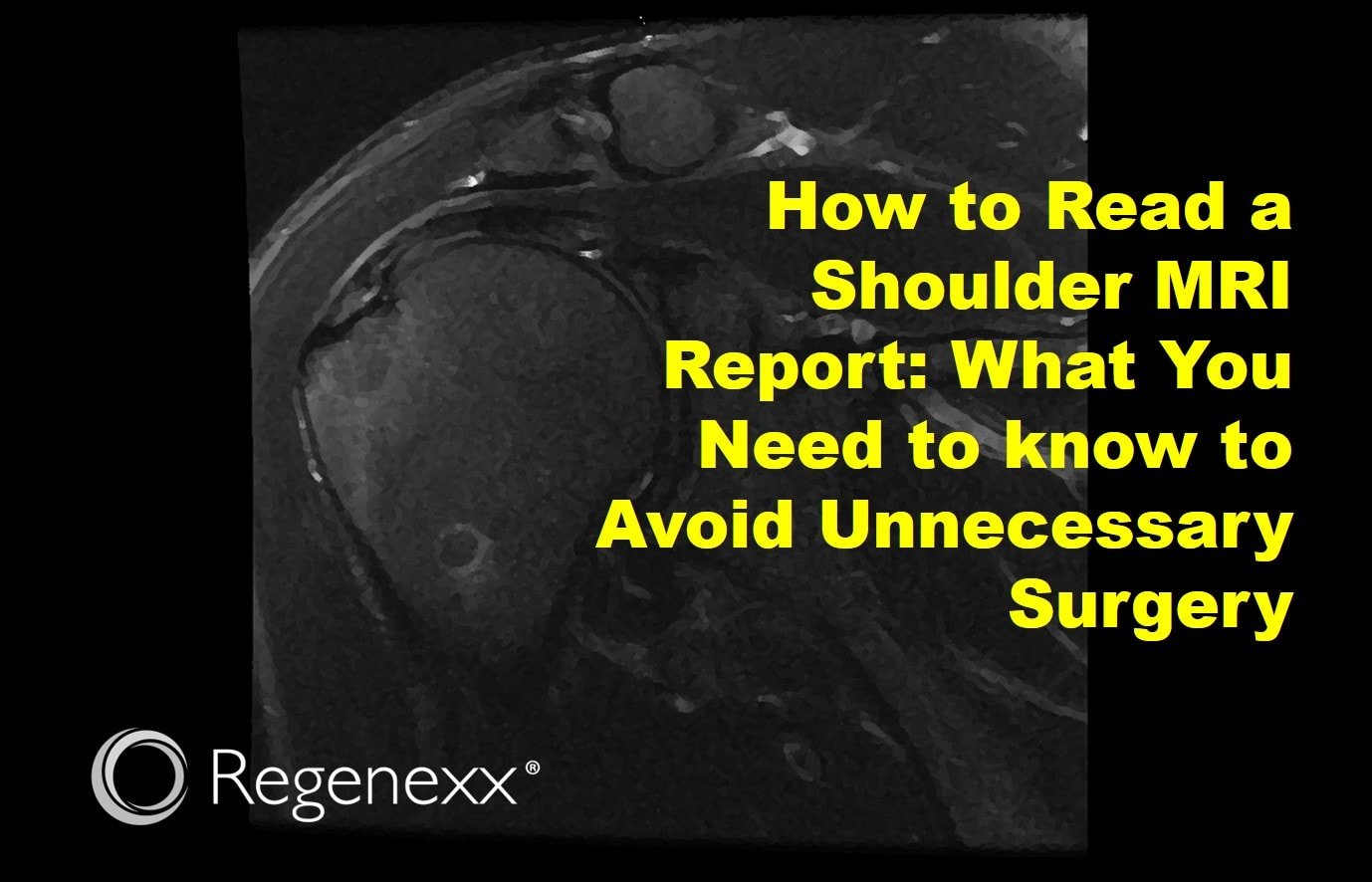
Impingement and rotator cuff tear
Frequently Asked Questions
What are the common indications for an MRI of the shoulder?
Doctors typically recommend an MRI of the shoulder to investigate various concerns such as persistent pain, weakness, or limited range of motion. Common conditions include rotator cuff tears, shoulder impingement, labral tears, and arthritis. MRI helps visualize soft tissues and cartilage, providing valuable information for diagnosis and treatment planning.
How can I interpret the findings of a shoulder MRI?
To interpret the findings of a shoulder MRI, look for specific structures such as the rotator cuff, labrum, and bursa. A normal MRI shows intact tendons, no signs of inflammation, and normal joint spaces. Abnormal findings might include tears, fluid accumulation, or bone spurs. It’s essential to discuss the results with a physician who can explain them in the context of your symptoms and medical history.
What role does contrast material play in shoulder MRI?
Contrast material enhances the visibility of certain tissues in an MRI. In shoulder imaging, physicians may use contrast to better visualize soft tissue structures, such as the labrum or to detect any tears in the rotator cuff. This can help differentiate between various conditions and improve the accuracy of the diagnosis.
What should I expect during a shoulder MRI examination?
During a shoulder MRI examination, a technician places you in the MRI machine and positions your arm to obtain clear images of the shoulder area. The procedure typically lasts 30 to 60 minutes. You will hear loud noises from the machine, but you can often listen to music through headphones. It’s important to remain still during the process to ensure high-quality images.
Can an MRI detect all shoulder issues?
An MRI is highly effective for diagnosing many shoulder issues, particularly those involving soft tissues. However, it may not detect certain conditions like fractures or subtle changes in bone unless accompanied by associated soft tissue findings. Complementary imaging techniques, such as X-rays or CT scans, may provide additional insights for a comprehensive evaluation.
Final Thoughts
Normal vs abnormal shoulder MRI results provide critical insights into shoulder health. A normal MRI shows no tears, inflammation, or structural anomalies, reflecting proper function and anatomy.
In contrast, an abnormal MRI reveals issues like rotator cuff tears, tendonitis, or joint degeneration, which can lead to pain and limited mobility.
Understanding these differences helps in diagnosing and treating shoulder conditions effectively, guiding patients towards appropriate interventions and recovery strategies. Evaluating MRI results is essential for optimal shoulder health management.